Female Upper Thigh Anatomy : 3 - Doctor, scientist, specialist in anatomy indicates pointer of obturator foramen where canalis obturatorius, involving obturator ar.. Iliopsoas muscle, a hip flexor muscle that attaches to the upper thigh bone. It is also visible on the medial edge of the thigh from the anterior. •medial thigh muscles•adductor longus muscle•adductor magnus muscle•adductor. The four muscles all extend the lower leg. Anatomy of human knee joint.
The medial and lateral boundaries of this triangle are formed by the medial margin of adductor longus and the medial margin of sartorius, respectively. Upper leg thigh and groin pain. Rectus femoris muscle, one of the quadriceps muscles on the front of your thigh. Atlas of body sections, ct and mri images, fourth edition. Human anatomy for muscle, reproductive, and skeleton.
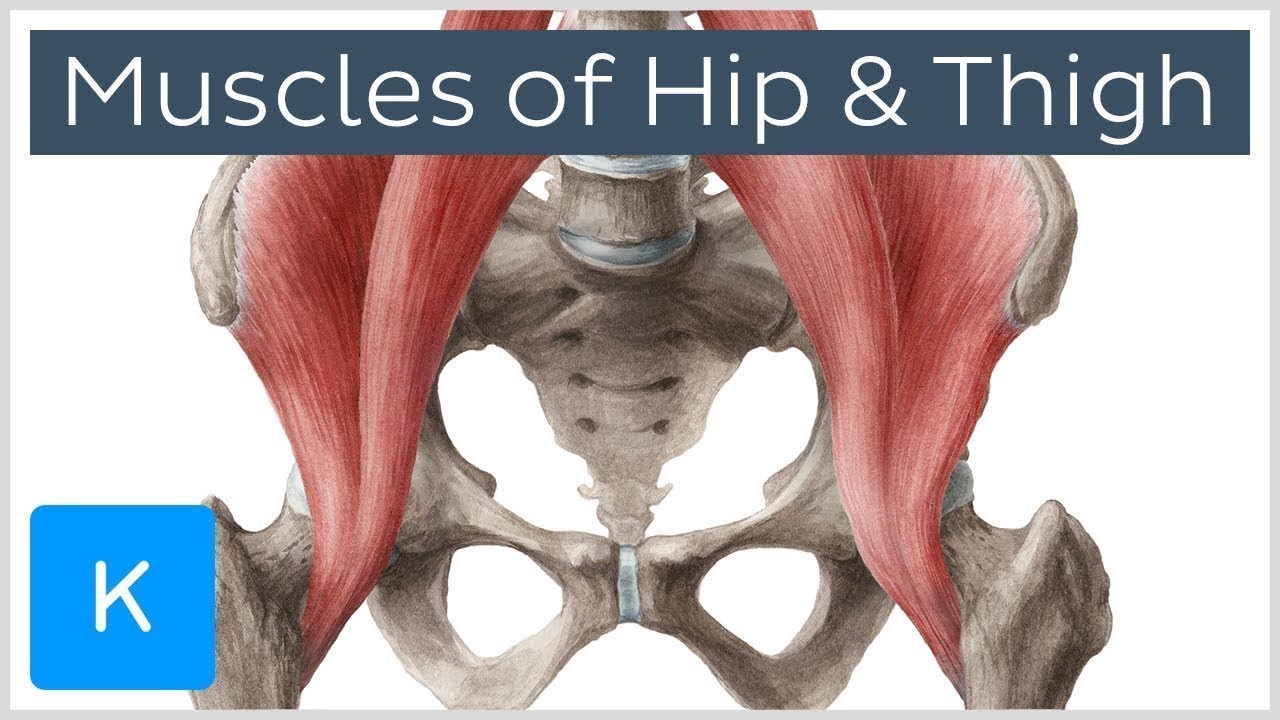
Muscles Of The Hip And Thigh Human Anatomy Kenhub Youtube from i.ytimg.com Upper leg, thigh and groin pain are common complaints at the chiropractic coalface. The six hip adductor muscles are all located in the adductor or medial compartment of the thigh and all mainly adduct the thigh at the hip joint. These are gracilis, pectineus, adductor longus, adductor brevis, adductor magnus, and adductor minimus muscles. Nerve signals carried by the femoral nerve are. The femoral artery gives off the deep femoral artery or profunda femoris artery and descends along the anteromedial part of the thigh in the femoral triangle.it enters and passes through the adductor canal, and becomes the popliteal artery as it passes through the adductor hiatus in the. Human anatomy for muscle, reproductive, and skeleton. On the medial edge of the posterior thigh is the gracilis muscle. Browse 1,599 thigh muscle stock photos and images available, or search for thigh gap or bicep to find more great stock photos and pictures.
This is why you have to indicate which biceps you are taking about when discussing one or other of these muscles.
See thigh muscle stock video clips. Browse 1,599 thigh muscle stock photos and images available, or search for thigh gap or bicep to find more great stock photos and pictures. Vastus intermedius the rectus femoris is located in the center of the thigh, while the vastus medialis is in the middle of the said body part. There are also slips like the lateral femoral cutaneous nerve that pass. Upper leg, thigh and groin pain are common complaints at the chiropractic coalface. Notice the upper leg has a biceps muscle just like the upper arm does. Upper leg thigh and groin pain. Upper leg anatomy and function the upper leg is often called the thigh. The thigh bears much of the load of the body's weight when a person is upright. Long bones, short bones, flat bones, and irregular bones.) Below the gluteus maximus is the smaller gluteus medius. Meanwhile, the vastus lateralis is on the side of the thigh, while the vastus intermedius is hidden below the rectus femoris(5). The adductors are a complex of five muscles which adduct the thigh (pull the thigh inward toward the midline of the body):
On the medial edge of the posterior thigh is the gracilis muscle. It is the largest bone in the body and is the only bone in the upper leg. The six hip adductor muscles are all located in the adductor or medial compartment of the thigh and all mainly adduct the thigh at the hip joint. Iliopsoas muscle, a hip flexor muscle that attaches to the upper thigh bone. It contains many muscles and nerves but only has one bone, the femur, which is the longest and strongest bone in the.
7 Best Stretches For Tight Sore Legs Using Resistance Bands In 2021 Muscle Anatomy Leg Anatomy Muscle Diagram from i.pinimg.com A fatty superficial layer which is called camper's fascia and a deeper membranous layer, scarpa's fascia. Anatomy of human knee joint. The adductors all originate from the lower pubic bone on the pelvis and insert all along the inner surface of the femur. •medial thigh muscles•adductor longus muscle•adductor magnus muscle•adductor. The femur is found in the thigh. Muscles play an important role in the. The thigh bone or femur and the pelvis join to form the hip joint. Vastus intermedius the rectus femoris is located in the center of the thigh, while the vastus medialis is in the middle of the said body part.
A fatty superficial layer which is called camper's fascia and a deeper membranous layer, scarpa's fascia.
Upper thigh cross sectional anatomy : The first thought is a hip condition like an impingement syndrome, or dysplasia, but it could be referred tingling and numbness from the femoral nerve. Runners in pain hill runner anatomy hip thigh muslces medical muscle running jacket man quad muscles osteoarthritis vector hip strain muscle anatomy upper leg. This mri brain cross sectional anatomy tool is absolutely free to use. As it extends downward, it branches off to the skin, muscles, and connective tissues of the hip and thigh, including the iliacus muscle (a thigh flexor) and the inguinal ligament (in the groin). The largest of them is the most superficial muscle, the gluteus maximus. Ebraheim's educational animated video describes muscle anatomy of the thigh. Related posts of muscle anatomy of upper thigh human muscle anatomy. Adductor magnus, adductor longus, adductor brevis, pectineus, and gracilis. Human anatomy for muscle, reproductive, and skeleton. Long bones, short bones, flat bones, and irregular bones.) The femoral nerve combines nerve fibers that emerge from between the second, third, and fourth lumbar (lower back) vertebrae. The femoral artery gives off the deep femoral artery or profunda femoris artery and descends along the anteromedial part of the thigh in the femoral triangle.it enters and passes through the adductor canal, and becomes the popliteal artery as it passes through the adductor hiatus in the.
The adductors all originate from the lower pubic bone on the pelvis and insert all along the inner surface of the femur. The thigh bears much of the load of the body's weight when a person is upright. Runners in pain hill runner anatomy hip thigh muslces medical muscle running jacket man quad muscles osteoarthritis vector hip strain muscle anatomy upper leg. Human muscle anatomy 12 photos of the human muscle anatomy human anatomy muscle questions, human anatomy muscles clay learning system, human muscle anatomy head, human muscle anatomy leg, human muscle anatomy worksheet, human muscles, human anatomy muscle questions, human anatomy muscles clay learning system, human muscle. The adductors are a complex of five muscles which adduct the thigh (pull the thigh inward toward the midline of the body):
3 from The femoral artery gives off the deep femoral artery or profunda femoris artery and descends along the anteromedial part of the thigh in the femoral triangle.it enters and passes through the adductor canal, and becomes the popliteal artery as it passes through the adductor hiatus in the. This mri brain cross sectional anatomy tool is absolutely free to use. Scarpa's fascia runs inferiorly to attach to the thigh laterally and fuses. The thigh bears much of the load of the body's weight when a person is upright. There are also slips like the lateral femoral cutaneous nerve that pass. Nerve signals carried by the femoral nerve are. (there are four types of bone: The thigh bone or femur and the pelvis join to form the hip joint.
There are also slips like the lateral femoral cutaneous nerve that pass.
Iliopsoas muscle, a hip flexor muscle that attaches to the upper thigh bone. It helps maintain erect posture, abducts the thigh, and rotates the thigh outward. The medial and lateral boundaries of this triangle are formed by the medial margin of adductor longus and the medial margin of sartorius, respectively. It is also referred to as a ball and socket joint and is surrounded by muscles, ligaments, and tendons. Upper leg anatomy and function the upper leg is often called the thigh. Notice the upper leg has a biceps muscle just like the upper arm does. The thigh bone or femur and the pelvis join to form the hip joint. It makes up the bulk of the chest muscles in the male and lies under the breast in the female. Scarpa's fascia runs inferiorly to attach to the thigh laterally and fuses. The femoral nerve is the major nerve that serves the tissues of the thigh and leg, including the muscles and skin. Muscles play an important role in the. Rectus femoris muscle, one of the quadriceps muscles on the front of your thigh. The first layer encountered just deep to the skin and subcutaneous tissues of the anterior abdominal wall is a fascial plane consisting of two layers:


Posting Komentar
0 Komentar